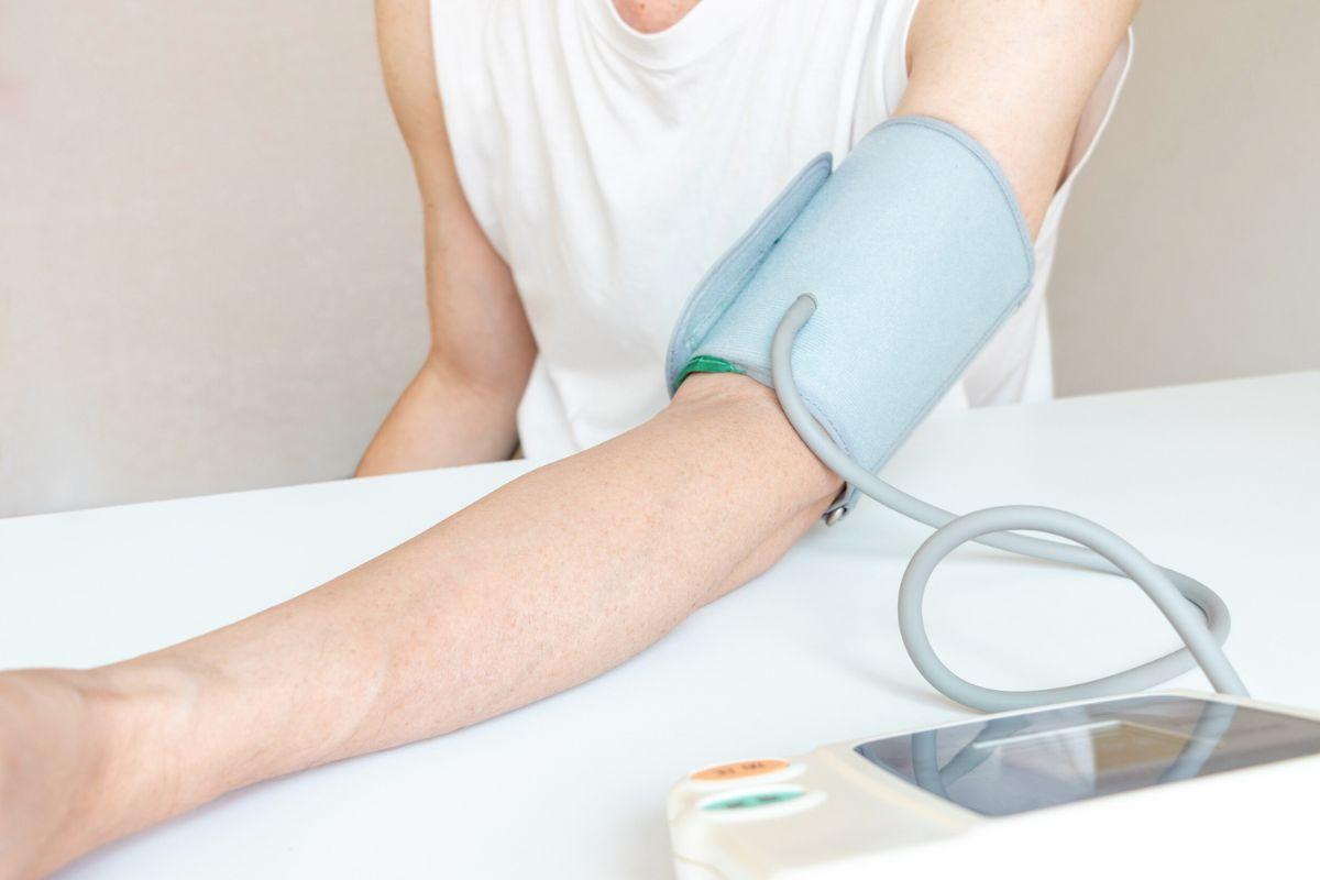
Being referred for a heart scan can feel worrying or confusing, especially if you’re unsure what the test involves or why it’s needed. The reassuring news is that both cardiac MRI and CT scans are safe, non-invasive procedures that give doctors highly detailed images of your heart and blood vessels.
These scans help identify problems early, guide treatment decisions, and give you peace of mind about your heart health. Doctify makes it easy to connect with trusted cardiologists who use advanced imaging to assess, monitor, and protect your heart.
Understanding Heart Imaging
Standard heart tests like ECGs and blood work are useful for spotting electrical or biochemical changes, but they can’t show what your heart actually looks like. That’s where imaging comes in.
While an ECG records your heart’s rhythm and electrical activity, cardiac imaging provides a clear picture of its structure, blood flow, and function. It allows doctors to identify problems that may not be visible on a standard heart tracing, such as narrowed arteries, damaged muscle tissue, or valve issues.
What is a Cardiac MRI?
A cardiac MRI (magnetic resonance imaging) uses powerful magnets and radio waves to create 3D images of the heart’s chambers, muscle walls, and blood flow – all without radiation.
This scan provides detailed information about how well your heart muscle is working and whether there are signs of scarring, inflammation, or congenital abnormalities. Doctors commonly use cardiac MRI to:
- Assess heart muscle strength after a heart attack or in cardiomyopathy.
- Detect scarring or inflammation, such as myocarditis.
- Evaluate heart valve function and congenital heart defects.
The test is painless and typically lasts between 30 and 60 minutes.
What is a Cardiac CT Scan?
A cardiac CT (computed tomography) scan uses low-dose X-rays to take rapid, cross-sectional images of your heart and surrounding blood vessels. Often, a special contrast dye is injected to highlight the arteries and help doctors assess blood flow.
Cardiac CT scans are commonly used to:
- Check for narrowed or blocked coronary arteries through coronary CT angiography (CCTA).
- Detect calcium build-up using a coronary calcium score.
- Map heart and vessel anatomy before surgery or other procedures.
The scan takes just 5 to 10 minutes and provides exceptionally clear images of your arteries, making it a powerful tool for assessing cardiovascular risk.
When Your Doctor Might Recommend These Tests
Your doctor may recommend a cardiac MRI or CT if you’re experiencing unexplained chest pain, breathlessness, palpitations, or abnormal ECG results. They’re also useful for:
- Monitoring known conditions such as hypertension, coronary artery disease, or heart failure.
- Assessing congenital heart defects or planning for surgery.
- Following up after treatment to evaluate recovery or response to medication.
These imaging tests give your care team the insight needed to make informed, precise treatment decisions.
How they compare: MRI vs CT
Both cardiac MRI and CT scans offer detailed insight into heart health, but they serve slightly different purposes.
- MRI provides superior soft-tissue detail, showing how the heart muscle moves and functions. It’s ideal for evaluating scarring or inflammation.
- CT is faster and particularly useful for visualising arteries and detecting blockages. It involves a small amount of radiation but delivers immediate, high-resolution images.
Your doctor will recommend the most appropriate test based on your symptoms, health history, and what they need to investigate.
What to expect during the scan
During a Cardiac MRI
You’ll lie comfortably inside the MRI scanner while images are taken. The machine makes a loud tapping or humming noise, but the test is painless. In some cases, a contrast dye is injected into your arm to enhance image clarity. You’ll need to remain still and occasionally hold your breath for short periods.
During a Cardiac CT
You’ll lie on a moving table that passes through the CT scanner. If contrast dye is used, you may feel a warm sensation briefly as it enters your bloodstream. The radiographer will give simple breathing instructions to help capture clear pictures. The entire process is typically over in under 10 minutes.
Risks and Safety
Both cardiac MRI and CT scans are considered very safe.
- MRI does not use radiation, but may not be suitable for people with certain pacemakers, metal implants, or severe claustrophobia.
- CT uses a small dose of X-rays, kept as low as possible for safety.
- In both tests, contrast dye reactions are rare and usually mild. Your medical team will check your kidney function and allergy history beforehand to ensure suitability.
After the test
Most people can return to normal activities immediately after their scan. If contrast dye was used, drinking plenty of fluids helps flush it from your system.
Your results will be reviewed by a radiologist and cardiologist, who will explain what the images show and whether any further action is needed. Follow-up may include lifestyle recommendations, medication adjustments, or additional investigations to monitor your heart health.
Conclusion
A cardiac MRI or CT scan provides an invaluable window into your heart’s health- helping doctors detect problems early, plan the best treatment, and prevent future complications. These tests are quick, safe, and highly informative, offering peace of mind through accurate diagnosis.
If you’ve been referred for heart imaging or would like to discuss your options, book an appointment through Doctify with a verified cardiologist who can guide you through the process and help you stay proactive about your heart health.
Find the right specialist for you. Doctify uses verified reviews so you can make the best decision for your healthcare.

Find the best cardiologists in the United Kingdom, or search for trusted experts globally:
- Cardiologists in Ireland
- Cardiologists in Australia
- Cardiologists in the United Arab Emirates
- Cardiologists in Germany
Medically Reviewed
Last reviewed on 28/10/2025